When it comes to health, early detection is often the key to effective treatment. For conditions like brain tumors, identifying warning signs as soon as possible can make a world of difference. Interestingly, your eyes may hold clues that could lead to the early detection of such serious conditions. Can an eye exam detect a brain tumor? This question has gained attention in recent years as researchers and medical professionals uncover the intricate connection between eye health and neurological disorders. While eye exams are primarily designed to evaluate vision and eye health, they can sometimes reveal signs of underlying systemic issues, including brain tumors.
During a routine eye exam, optometrists and ophthalmologists examine not just your vision but also the structures of your eyes. The retina, optic nerve, and blood vessels are all carefully inspected for abnormalities. In some cases, these professionals notice signs that may point to problems beyond the eyes. For instance, changes in the optic nerve or unexplained vision loss could indicate pressure or swelling in the brain, which might be caused by a tumor. While an eye exam alone cannot diagnose a brain tumor, it can serve as an important first step in identifying potential red flags that warrant further investigation.
Understanding the relationship between eye exams and brain tumors is crucial for both patients and healthcare providers. Many people may not realize that their eyes can act as a window to their overall health. By staying informed about the signs and symptoms that could arise during an eye exam, individuals can take proactive steps toward their well-being. In this article, we will explore how eye exams can help detect brain tumors, the specific signs optometrists look for, and what steps to take if abnormalities are found. Let’s dive into the details and uncover the fascinating ways our eyes can provide critical insights into our health.
Read also:Astro Kpop Band The Meteoric Rise Of A Stellar Group
Table of Contents
- How Do Eye Exams Work?
- What Signs During an Eye Exam Could Indicate a Brain Tumor?
- What Is the Connection Between the Eyes and the Brain?
- Which Types of Brain Tumors Can Affect Vision?
- What Diagnostic Tools Are Used After an Eye Exam?
- Why Are Regular Eye Exams Important for Early Detection?
- When Should You See a Doctor About Vision Changes?
- Can an Eye Exam Detect a Brain Tumor? Summing It All Up
How Do Eye Exams Work?
Eye exams are comprehensive evaluations that go far beyond simply checking whether you need glasses. They involve a series of tests designed to assess the health of your eyes and detect any abnormalities. During a typical eye exam, an optometrist or ophthalmologist will first review your medical history, asking about any vision problems, eye discomfort, or family history of eye diseases. This step is crucial because it provides context for the rest of the examination and helps the doctor identify potential risk factors.
Next, the doctor will perform a series of tests to evaluate different aspects of your eye health. These may include a visual acuity test to measure how well you can see at various distances, a refraction test to determine your prescription for glasses or contact lenses, and a slit-lamp examination to inspect the front and back of your eye. One of the most critical parts of the exam is the dilated eye exam, where drops are used to widen your pupils. This allows the doctor to examine the retina and optic nerve in detail, which is where signs of systemic conditions like brain tumors might be detected.
While the primary goal of an eye exam is to ensure your vision is clear and your eyes are healthy, it can also reveal underlying health issues. For example, changes in the optic nerve, such as swelling (a condition known as papilledema), can indicate increased intracranial pressure, which may be caused by a brain tumor. Similarly, unexplained vision loss or double vision could point to neurological problems. Although an eye exam cannot diagnose a brain tumor on its own, it can prompt further investigation and lead to early detection of serious conditions.
What Signs During an Eye Exam Could Indicate a Brain Tumor?
During an eye exam, certain signs and symptoms may raise red flags about the possibility of a brain tumor. One of the most telling indicators is papilledema, or swelling of the optic nerve. This condition occurs when there is increased pressure inside the skull, which can compress the optic nerve and cause it to swell. Papilledema is often painless, but it can lead to vision problems if left untreated. Optometrists and ophthalmologists are trained to recognize this condition during a dilated eye exam, making it a critical tool for early detection.
Are There Other Vision-Related Symptoms to Watch For?
Aside from papilledema, other vision-related symptoms may signal a potential brain tumor. For instance, sudden or unexplained vision loss in one or both eyes could indicate pressure on the optic nerve or visual pathways in the brain. Double vision, also known as diplopia, is another warning sign. This occurs when the eyes are unable to align properly, often due to pressure or damage to the cranial nerves that control eye movement. Additionally, patients may experience blurred vision or difficulty focusing, which can be caused by swelling or compression in the brain.
Could Changes in the Retina Be a Clue?
Yes, changes in the retina can also provide valuable clues. For example, retinal hemorrhages or abnormal blood vessel patterns may suggest underlying systemic issues, including brain tumors. These changes are typically detected during a dilated eye exam, where the doctor uses specialized instruments to examine the back of the eye. While these signs alone do not confirm the presence of a brain tumor, they can prompt further diagnostic testing, such as imaging scans, to rule out or confirm the diagnosis.
Read also:Ryans World Controversy Unpacking The Issues Surrounding The Popular Kids Channel
It’s important to note that these symptoms are not exclusive to brain tumors and can be caused by a variety of other conditions, such as migraines, glaucoma, or diabetes. However, when they occur in combination or are accompanied by other neurological symptoms like headaches, nausea, or seizures, they should be taken seriously. Early detection through an eye exam can lead to timely intervention, improving the chances of successful treatment.
What Is the Connection Between the Eyes and the Brain?
The eyes and the brain are intricately connected, forming a complex network that allows us to perceive the world around us. The optic nerve, which acts as a direct pathway between the eyes and the brain, plays a crucial role in this relationship. This nerve carries visual information from the retina to the brain, where it is processed and interpreted. Because of this close connection, any disruption in the brain can often manifest as changes in vision or eye health, making the eyes a valuable diagnostic tool for detecting neurological conditions.
One of the reasons the eyes are so effective at revealing brain-related issues is their accessibility. Unlike the brain, which is protected by the skull and requires advanced imaging techniques to examine, the eyes can be directly observed during an eye exam. This allows doctors to detect abnormalities such as swelling, bleeding, or damage to the optic nerve, all of which could indicate problems in the brain. For example, increased intracranial pressure—a common symptom of brain tumors—can cause the optic nerve to swell, a condition known as papilledema. By examining the eyes, doctors can gain insights into what might be happening inside the skull.
Furthermore, the eyes are controlled by cranial nerves, which originate in the brain. These nerves govern functions such as eye movement, pupil dilation, and focusing. Any damage or pressure on these nerves can lead to symptoms like double vision, drooping eyelids, or difficulty moving the eyes. These symptoms can serve as early warning signs of a brain tumor or other neurological disorders. By understanding the connection between the eyes and the brain, healthcare providers can use eye exams as a non-invasive way to screen for potential issues and guide patients toward further diagnostic testing when necessary.
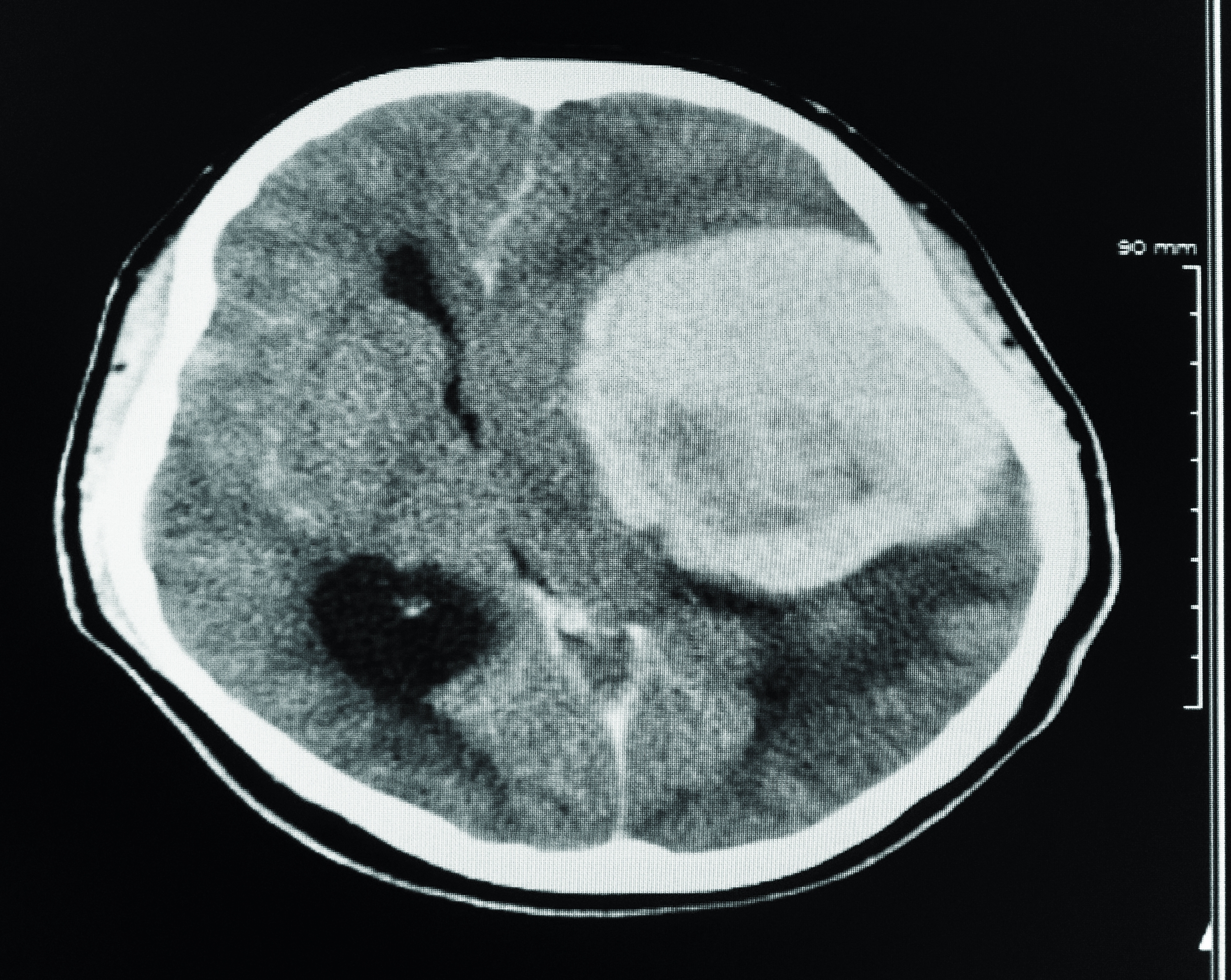
Which Types of Brain Tumors Can Affect Vision?
Not all brain tumors affect vision, but certain types are more likely to cause visual disturbances due to their location or size. One of the most common culprits is a meningioma, a tumor that arises from the meninges, the protective layers surrounding the brain and spinal cord. Meningiomas often develop near the optic nerve or in areas that control eye movement, leading to symptoms like blurred vision, double vision, or even vision loss. Because these tumors grow slowly, symptoms may develop gradually, making them harder to detect without a thorough eye exam.
Pituitary adenomas are another type of brain tumor that can impact vision. These tumors originate in the pituitary gland, a small structure located at the base of the brain. As they grow, they can press on the optic chiasm, the point where the optic nerves from each eye intersect. This pressure can cause a specific type of vision loss known as bitemporal hemianopia, where the outer halves of the visual field in both eyes are affected. Patients may notice difficulty seeing objects on the sides or trouble reading, which can prompt them to seek medical attention.
Gliomas, tumors that arise from glial cells in the brain, can also affect vision depending on their location. For example, a glioma in the occipital lobe—the part of the brain responsible for processing visual information—can lead to partial or complete vision loss. Similarly, tumors in the brainstem or cerebellum may interfere with the cranial nerves that control eye movement, resulting in double vision or difficulty focusing. While these tumors are less common than meningiomas or pituitary adenomas, they can cause significant visual disturbances that are detectable during an eye exam.
What Diagnostic Tools Are Used After an Eye Exam?
If an eye exam reveals signs that could indicate a brain tumor, the next step is to use advanced diagnostic tools to confirm or rule out the diagnosis. One of the most common methods is magnetic resonance imaging (MRI), which provides detailed images of the brain and surrounding structures. An MRI can identify the location, size, and type of tumor, as well as any associated swelling or compression of the optic nerve. In some cases, a contrast agent is used to enhance the clarity of the images, making it easier to detect abnormalities.
Could a CT Scan Be Necessary?
Yes, a computed tomography (CT) scan may also be used, especially if an MRI is not feasible. While CT scans are not as detailed as MRIs, they can still provide valuable information about the presence of a tumor and its impact on surrounding tissues. CT scans are particularly useful for detecting calcifications or bone changes, which may not be as visible on an MRI. Both imaging techniques are essential for confirming the findings of an eye exam and guiding treatment decisions.
What About Additional Tests?
In addition to imaging, doctors may order other tests to gather more information. For example, a lumbar puncture, or spinal tap, may be performed to analyze cerebrospinal fluid for signs of tumor cells or inflammation. Blood tests can also help identify hormonal imbalances caused by pituitary tumors. These diagnostic tools, combined with the initial findings from an eye exam, provide a comprehensive picture of the patient’s condition and help determine the best course of action.
Why Are Regular Eye Exams Important for Early Detection?
Regular eye exams are more than just a routine checkup; they are a vital tool for maintaining overall health. While most people associate these exams with correcting vision problems, they also play a crucial role in detecting systemic conditions like brain tumors. Early detection is key to successful treatment, and eye exams can often reveal warning signs before symptoms become severe. For example, subtle changes in the optic nerve or retina may not cause noticeable symptoms but can be identified by a trained eye care professional during a routine exam.
One of the reasons regular eye exams are so effective is their ability to catch problems early. Many brain tumors develop gradually, and symptoms like vision changes or headaches may be dismissed as minor inconveniences. However, an optometrist or ophthalmologist can spot abnormalities that might otherwise go unnoticed. For instance, papilledema, or swelling of the optic nerve, is often painless and may not cause immediate vision loss, but it can be detected during a dilated eye exam. Identifying such signs early can lead to timely intervention and improve treatment outcomes.
Moreover, regular eye exams are particularly important for individuals with risk factors for brain tumors, such as a family history of neurological conditions or previous head injuries. By scheduling annual or biannual eye exams, these individuals can monitor their eye health and ensure any changes are addressed promptly. Even for those without known risk factors, routine exams provide peace of mind and serve as an opportunity to discuss any concerns with a healthcare professional. In short, regular eye exams are a simple yet powerful way to safeguard your health and detect potential issues before they become serious.
When Should You See a Doctor About Vision Changes?
Knowing when to seek medical attention for vision changes can make all the difference in diagnosing and treating serious conditions like brain tumors. While occasional vision blurriness or eye strain is common, certain symptoms warrant immediate evaluation by a healthcare professional. For instance